
The following figure is presented here at reduced scale (resolution)to accommodate a browser. It is completely inadequate to make the desired point. A larger scale version is available in Chapter 3 in the Download Files area reached from the Site navigation bar.
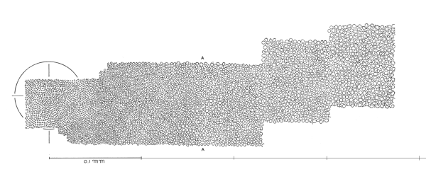
Topography of the human retina (Section taken through the inner segments)
[from Section 3.2]
There are virtually no figures in the literature showing the size and density of the Outer Segments of the human retina as a function of position over a significant fraction of the retina. Only a very few images exist showing a significant portion of the retina at the level of the Inner Segments. Others show cross-sections of the photoreceptor cells taken at more proximal locations within the retina. Of the few large area maps, that due originally to Pirenne is probably the best available. It shows the cross-section of the retina through the Inner Segments. Most authors have truncated his imagery for convenience in printing. Even Pirenne published his original figure in sections. The above figure reassembles those sections to focus on the fact that the size and density of the Inner Segments, taken here as a proxy for a true map of the Outer Segments, does not vary significantly.
Whereas the spatial resolution of the human eye decreases by more than an order of magnitude within a few degrees of the line of sight (and at least two orders at ten degrees from the line of sight, the density of the Inner Segments decreases by less than a factor of 5 between the center of the fovea and the extremes of the retina.
The diameter of the Inner Segments of the photoreceptors are essentially of constant diameter throughout the human retina except for the very small region known as the foveola.
The nominal diameter of the Outer Segments is found to be 2.0 microns throughout the majority of the human retina.
The above features are discussed in detail in Chapter 3.
Return to the website home page