The following figure is presented here at reduced scale (resolution)to accommodate a browser. A larger scale version is available in Chapter 4 in the Download Files area reached from the Site navigation bar.

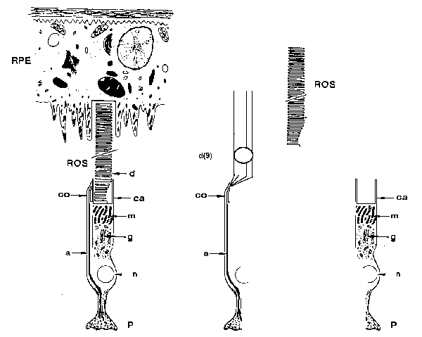
Topography of the photoreceptor cell of vision (including an exploded view)
[from Section 4.2]
The figure on the left shows a complete photoreceptor cell in its normal environment. A photoreceptor cell is classed as a neuro-secretory type cell of the Neural System. The receptor Outer Segment (ROS) is shown forshortened because of the very high length to diameter ratio of an Outer Segment, typically 25:1.
The ROS is shown extending into a "cup" of the retinal pigment epithelium (RPE) where the ROS is disassembled and its materials recycled as part of the process of phagocytosis.
The lettering represents: a=axon of the neuron, ca=calyx, co=colax, d=disks, d(9)=the nominally nine dendrites (also known as microtubules in morphology), g=Golgi apparatus, m=mitochondria, n=nucleus, P=pedicel.
The nine dendrites are arranged in a ring with each dendrite placed in a furrow of the disk stack formed by the extrusion process performed by the calyx, a part of the Inner Segment of the cell. following secretion of the protein opsin into a strip within the limited volume of the calyx, three events occur. First, the strip is repeatedly bent until it breaks into a series of roughly square pieces. Second these pieces are formed into a stack. Third the stack is formed into a circular column by the extrusion die at the end of the calyx.
The active elements of the photoreceptor cell, the Activas, are located within the individual dendritic structures of the cell and just inside the cell body near the colax (also known as the ciliary collar in morphology).
Following extrusion, the disk stack forming the main structure of the Outer Segment leaves the photoreceptor cell environs and is immersed in the Inter-Photoreceptor cell Matrix (IPM) until is arrives at the phagocytosis cup of the RPE. The disk stack is not surrounded by a cell membrane while it is in the IPM although it is coated with a variety of nutrients supplied from the RPE layer. In humans, it takes a given disk about 12 weeks to travel from the point of formation to its point of phagocytosis.
The nucleus of the cell plays no role in the functional aspects of the neuron. The Golgi apparatus and the mitochondria provide the protein, opsin, that is secreted by the cell and used to form the disks.
The above features are discussed in detail in Chapter 4.
Return to the website home page