
The following figure is presented here at reduced scale to accommodate a browser.A larger scale version is available in Chapter 7 in the Download Files area reached from the Site navigation bar.
This summary figure is complex and can not be described completely here. Section 7.4.1 of the text needs to be referred to for a full discussion. The figure stresses the important structural dynamics of the living retina as a function of time.
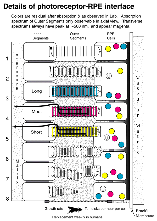
The Photoreceptor-RPE interface in the human Visual System
[from Section 7.4.1]
Row 1 illustrates the formation of disks within the calyx of the photoreceptor cell, their formation of a stack of disks known as the Outer Segment, and the disassembly of these disks by the process of phagocytosis when they have reached the RPE cells. As noted at the bottom of the figure, disks are generated at a rate of approximately one per hour and the life expectancy of a disk is approximately 12 weeks in humans.
Row 2 shows a situation similar to row 1 except the disks have become coated with chromophoric material transported from the RPE cells through the IPM. The photoreceptor in row 2 is still not functional.
Row 3 shows the addition of electrostenolytic material surrounding and between the disks. This material provides the electrical power required to operate the dendrites of the photoreceptor cells.
Rows 4 and 5 show the presence of the dendrites (microtubules) in the furrows of the Outer Segments and their progression through the colax and into the Inner Segment of each cell.
Rows 3, 4 and 5 show separate spectral absorption channels. The colors shown are the nominal complement of the colors actually absorbed. e. g., the color of the Outer Segment observed by reflection when looking at the segment through the pupil of the eye.
Rows 6, 7 and 8 illustrate three pathological conditions. Row 6 shows the shearing of an Outer Segment without displacement due to a retinal tear. Except for possible damage to the dendrites of the cell, the photoreceptor cell remains fully intact. The portion of the Outer Segment adjacent to the photoreceptor cell may also remain functional. Following the tear, new disks will continue to be produced. This will force the old disks to move toward the RPE cells and eventual phagocytosis. The result is a self repairing situation.
The situation in Row 7 is more difficult, the displacement of the disks at the tear may cause complications in the repair process. This condition could lead to permanent separation of the retina from the RPE cell layer. If the tear involves rupture of the IPM space, other biologicals might enter the IPM. These materials could interfere with the complex electrochemistry existing in this space and result in permanent damage.
Row 8 symbolizes the formation of a new photoreceptor cell and Outer Segment. During the initial filling of the calyx with the protein opsin, it is possible that undersize disks could be formed. This could result in an Outer Segment that initially appears conical in shape. However, this is an unsustainable growth situation. As the volume within the calyx increases, the calyx will expand to its nominal limit. After that point, all new disks will be of the same nominal diameter. There is no plausible way to generate a conically shaped Outer Segment over a prolonged period of time in the presence of a continual transit of disks from the Inner Segment of the photoreceptor cell to the RPE.
Return to the website home page