
 Complete Circuit Diagram of the Visual System[from Section 11.6.4]
A larger scale version is available for download in the Download Files area reached from the Site navigation bar.
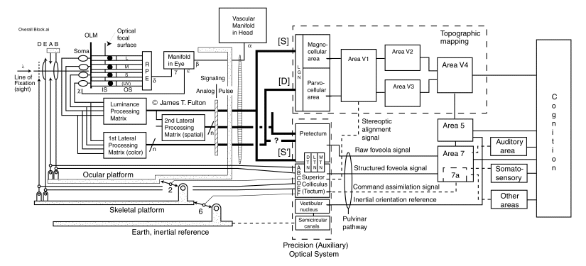
This Figure is the culmination of a great deal of analysis and is necessarily complex. It is believed to be the most complete block diagram of the visual system in existence. It can be best understood by exploring it in sections.
The upper left quadrant shows the eye of a visual system mounted on a multi-degree of freedom servo platform formed by the ocular and skeletal muscles. Light is seen projected through a number of optical elements before reaching the Optical focal surface. While this surface is highly curved in the real world, it is shown flat here for convenience. The adjustable optical elements shown consist of the iris (A), the Lens (B), the eye lid (D) and an auxiliary eye lid that is sometimes used as a nictating lens (E). The cornea (C) is not shown in this figure because it has a fixed optical power and is not subject to change by the servo system. There is also a field lens that is not shown explicitly. It is formed by the variable thickness of the neural tissue of the retina. Note also the black dots shown to the right of the Optical focal surface. These dots represent the "ellipsoids" found within many Inner Segments of the photoreceptor cells.
The Outer Segments of the photoreceptor cells are shown to the right of the ellipsoids. There are four varieties of these Outer Segments. Depending on the particular chromophore deposited on the disks of the segments, four individual spectral characteristics are obtained, the ultra-violet, the short, the middle, and the long wavelength regions of vision. It will be stressed elsewhere that the generic animal eye is a tetrachromatic eye. The human eye is restricted to trichromatic operation by environmental considerations.
The inner segments and the soma of the cells are shown on the left. The axons of these cells pass out through the illuminated face of the light sensitive part of the retina and into the neural portion shown below the photoreceptors. There are four primary signal processing matrices in the retina. Only three are shown explicitly.
Also shown in the upper left quadrant is a very important additional element. This is the vascular manifold of the eye. This manifold provides two different hydraulic paths to the retina. One feeds the neural portion of the retina. The second path feeds the Retinal Pigment Epithelial cells and the sclera of the eye. The hydraulic manifold and all of the elements discussed above, except the two eyelids reside on the ocular platform that is mechanically controlled by the ocular muscles. All of the neurons serving the eye (both afferent and efferent) all are contained within the optic nerve connecting the ocular globes to the brain. The optic nerve is shown as the large lens shaped structure to the right of the ocular platform.
Note the parallel connections of the luminance matrix and the 1st lateral matrix with the photoreceptors. This is very important in the architecture of the eye. Whereas only one type of signal path emanates from the luminance matrix, the luminance signal path labeled R in this work, there are multiple signal paths emanating from the 1st and 2nd lateral matrices. The 1st lateral matrix creates up to four distinct types of signal paths, but only two in long-wavelength trichromats such as humans. These paths are labeled the N-, O-, P- & Q- signal paths. The N- path prepares and transmits polarization information in animals, primary arthropods capable of using this information. The other paths derive a difference signal from pairs of spectrally sensitive photoreceptor channels. The O-channel represents the difference between the short wavelength (S-) spectral channel and the UV-channel. The P-channel represents the difference between the S- and M-channel, and the Q-channel represents the difference between the M-channel and the L-channel. Thus, the n next to the slash line indicates that multiple signal channels emanate from the 1st lateral matrix.
Multiple signals also emanate from the 2nd lateral matrix. This matrix is not used to a significant degree in humans. However, it is more important in other animals, particularly predators.
After the raw spectral signals are processed by the matrices and passed through the ganglion cells, they are distributed to the brain as shown on the right of the figure. The brain can be described in terms of a great variety of functional, cartographic and other characteristics. Here, the functional characteristics are of primary importance but the cartographic locations defined by morphology provide a convenient physical reference.
The upper right quadrant shows the portion of the brain most heavily studied to data. It is known to incorporate a degree of spatial mapping that correlates with the image of the scene impressed on the retina. However, this correlation is reduced for the higher V#'s in the dashed box. The signal paths marked (S) entering that box are the summation or luminance signals of vision. The signal paths marked (D) represent the difference signals, Primarily P- and Q- in the case of the human.
The signal paths marked (S') do not go to the topographically mapped portion of the brain. They are the spectral signals from the foveola. They go directly to the pretectum, a part of the Precision Optical System (POS) of the brain. The POS is the central processing unit of the main servomechanisms controlling the motions of the eyes and the head relative to the skeleton. This system processes the signals from the foveola to produce two precision output signals. One set of signals is used to control the neural centers below the pretectum that generate oculomotor control signals. The other set of signals consist of highly specific signals about the geometry of the image projected on the foveola. These signals cover a very small spatial area of the scene. They are labeled the raw foveola signals. These signals are passed directly to the area labeled variously in the literature as area 5 or area 7 of the cortex. These signals do not pass through area 17, 18,& 19 (roughly V1, V2 & V3, and V4).
The fact that the raw foveola signals do not pass through areas 17, 18,& 19 provides a time advantage in processing that can be significant. The advantage is measured in milliseconds.
A variety of additional features are shown in this figure that are discussed in detail in Chapters 11 and 15.