
The following figure is presented here at reduced scale (resolution)to accommodate a browser. A larger scale version is available in Chapter 8 in the Download Files area reached from the Site navigation bar.
There is an architecture more fundamental to the neural system than the neuron itself. This architecture is illustrated below. The neural system consists of a large array of unidirectional signaling channels. These channels may branch and combine. However, the basic architecture is very simple.
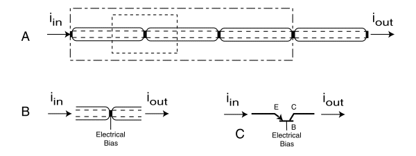
Fundamental architecture of the neural system
[from Section 8.4]
The fundamental signal path in the neural system consists of a series of electrolytic conduits connecting active electrolytic devices that can be considered as signal amplifiers (in the simplest connotation of that word). The system is entirely electronic in character and there is no requirement for chemical substances to flow along or between the conduits for signaling purposes. On the other hand, there is a need for materials to flow along the conduits, in a secondary role related to the metabolism and growth of the biological system. This requirement is discussed elsewhere in the text.
In A, the basic conduit is shown as the area between the parallel dashed lines. The area outside of these lines is involved with the biological functions of the neural system. Within these lines, the fundamental neurological process is the transmission of a signal in the form of an electronic current. As a practical matter, the signal must be amplified at discrete intervals along the length of the channel to insure an adequate signal amplitude at the output terminal. This amplification is performed by separating the overall channel into a series of shorter channels joined together in a unique manner.
B shows this method of "junction formation" without any detail. The important points to note are that the outer walls of the individual conduit sections are simple biological membranes that act as electrical diodes that are normally biased electrically to be non-conductive. There is negligible conduction of current through these walls except at the junctions.
At the junctions, three separate electrical potentials are found; the potential of the pre-junction conduit, the potential of the post junction conduit, and the potential of the surrounding medium. By juxtaposing the walls of the two conduits at a specific distance and thereby controlling the nature of the substance between them (to be explained in the text), a unique situation arises. "Transistor action" is obtained. This is the same transistor action found in man made devices. Obtaining this action in a biological system is covered by a United States Patent. The resulting situation is shown in C using standard electrical symbols and notation. The medium found between the two conduit walls is labeled the base. The pre-junction wall is labeled the emitter and the post junction wall is labeled the collector (of the charge passing through the junction).
This basic structure can be modified in a large variety of ways. However, in the simplest way, the current collected by the collector can be more than 99.9% of the current emitted by the emitter. Thus, the loss of signal current due to transmission through such a junction is negligible. In addition, the transmission time is extremely short because of the thickness of the junction, less than 100 Angstroms, ( or 10 nanometers).
It is important to understand the relationship between this fundamental signaling mechanism within the concept of a neuron. This is shown in the RELATIONSHIP BETWEEN THIS FUNDAMENTAL FUNCTION AND THE NEURON.
The above features are discussed in detail in Chapter 8.