
A larger scale version appears in Chapter 15 and is available for download in the Download Files area reached from the Site navigation bar.
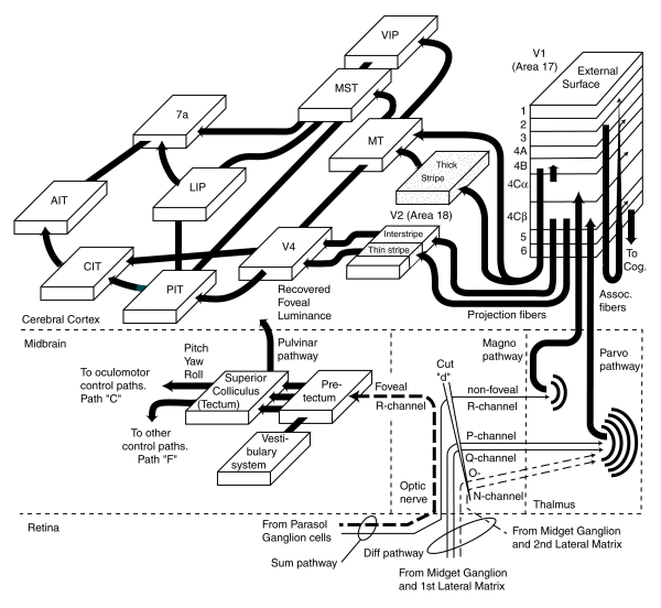
This figure illustrates how sensory information is fed to the higher perceptual areas of the cortex for evaluation via a series of feature extraction engines. The nomenclature is primarily that of the morphologist and electro-physiologist.
Note the two functionally unique entry points for visual signals reaching the cortex. The left entry point is for precision information from the foveola. The right entry point via the thalmus is for the less precision signals.
The signals transmitted from the mid-brain to the cortex appear to be primarily luminance (R-channel)signals. The signals transmitted from the thalmus to the cortex appear to include luminance and chrominance signals.
The signals transmitted from the Superior Colliculus and Pretectum are only recorded within the cortex in a vector format. They are not recorded within the cortex in a spatial context. Only the signals transmitted to the cortex via the thalmus are recorded initially in a spatially coherent manner. Even this mapping is lost in the areas forward of area 18.
Note the number of cross-connections of the various engines labeled within the cortex. The list of engines is only a small fraction of the total number of engines present and the number of interconnections shown is an even smaller number of the total number of connections involved. This pattern of interconnection is indicative of a "star" interconnection architecture.
The complete discussion of this figure is found in Section 15.2 of Chapter 15.