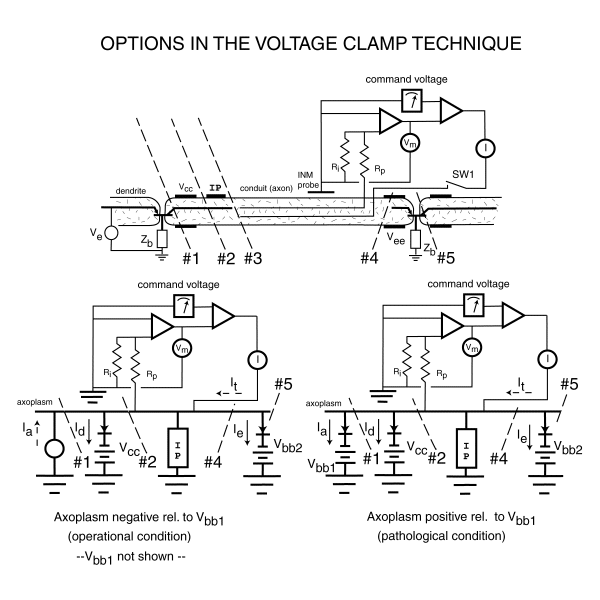
To employ the voltage clamp technique properly, it is mandatory that the investigator determine precisely where, and how, he interrupts the intrinsic neural path. The following figure illustrates five different important locations. Disecting the cell at each of the locations indicated results in a different experiment with different recorded results. To provide perspective, the locations #1 through #3 are frequently only a few microns apart. In the case of the giant squid axon, they may be as much as a millimeter apart.

THE SCHEMATIC OF THE VOLTAGE CLAMP TECHNIQUE [from Section 10.10.4]
The upper part of the above figure defines the test configuration and the morphology of the cell. The lower part of the figure defines the test configuration and the electrical topology of the neuron.
The lower two panels show the equivalent topology of the cell and the test configuration. It is important to note that the impedance of the unviolated axolemma is so high that neither its shunt resistance or shunt capacitance play a significant role in the experiment.
The lower left shows the nominal biological condition with the axoplasm negative with respect to both the surrounding fluid matrix and the voltage supply, Vbb1 connected to the base of the left-most Activa. Under this condition, the impedance of the left-most Activa is nominally infinite with respect to the rest of the circuit.
The lower right figure shows the pathological, although frequently presented, condition where the axoplasm has been forced to be positive with respect to the surrounding matrix and Vbb1. Under this condition, the impedance of the left-most Activa is now that of a low impedance diode in series with a small voltage.
The difference in performance of the "neuron" under these two conditions is dramatic. The difference is due to the operating conditions applicable to the left-most Activa and has nothing to do with the axolemma of the cell, which is passive.
Return to the website home page