Vitamins were recognized for their importance in good health beginning in the early 1900's. The role of Vitamin A was recognized for its importance in a number of body functions, but particulary for its importance in vision. Wald first showed that an animal deprived of Vitamin A soon became permanently blind.
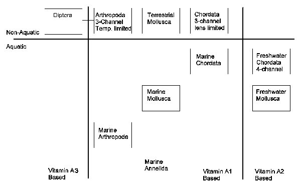
During Wald's experiments, it soon became apparent that there was more than one form of Vitamin A. It was soon established that one form was shared among all marine and other animals with a system based on salt water. This form became known as Vitamin A1. A second form was found in freshwater fish. It became known as Vitamin A2.
Recently, it has been found that large numbers of animals employ a third type of Vitamin A. This form appears to be common among animals that survive on carion. It appears that the carotenes, used by most animals to form Vitamin A, break down easily as the plant containing them decays. There are some carotenes that are more resistant. It is these carotenes that act as a source of this third type of Vitamin A, Vitamin A3. Vogt has found the visual systems of a wide variety of animals depend on Vitamin A3. The largest group are the two-winged flies of the Order Diptera and the moths of the Order Lepidoptera. However, Vitamin A3 has also been found in the eyes of crustaceans.
The eyes of Euryhaline fish, that transition from freshwater to seawater, are found to contain a mixture of Vitamin A1 and Vitamin A2. The percentage of each can be correlated to the recent travels of the animal. Similarly, the eyes of the flies in the Order Odonata are known to contain both Vitamin A1 and Vitamin A3. Research has only begun in the area of taxonomy related to Vitamin A. It is likely that many more discoveries are yet to appear.
The appearance of the different forms of Vitamin A can be illustrated using the following figure.

Chapter 1 of PROCESSES IN BIOLOGICAL VISION discusses the phylogenic role of Vitamin A in vision more completely. It also contains highr quality art than can be supported by the Web. It can be downloaded from the colorvision website.
The three prototypical forms of Vitamin A are shown in the figure. Vitamin A1 has long been known by the chemical name retinol. This material is instantly recognized by its chemical structure. It consists of a b-ionone ring attached to a conjugated chain of hydrocarbons terminating in an alcohol group.

These chemical formula are shown slightly differently than in most texts. This is to highlight the role of these materials as the chromogens of vision. By the addition of another oxygen to any of these molecules at the top of the stem associated with carbon number 5, the vertical stem at the upper left of each molecule, the material is converted from a retinene, the family name including the retinols, to a retinIne, the family name of the chromophores of vision. There are four individual chromophores with the name Rhodonine. Rhodonine(5) described above is sensitive in the red. Rhodonine(7) is sensitive in the green and Rhodonine(9) is sensitive in the blue. Rhodonine(11) is sensitive in the ultraviolet and is used primarily by the insects and other members of the phylum Arthropoda. Additional data on the chromophores of vision are available on the PROCESSES IN BIOLOGICAL VISION website.
Citations
Isler, O. (1971) Carotenoids, NY: Halsted Press, Div. of John Wiley & Sons
Vogt, K. (1989) In Stavenga, D. & Hardie, R. Facets of Vision, NY: Springer-Verlag, Chapter 7.