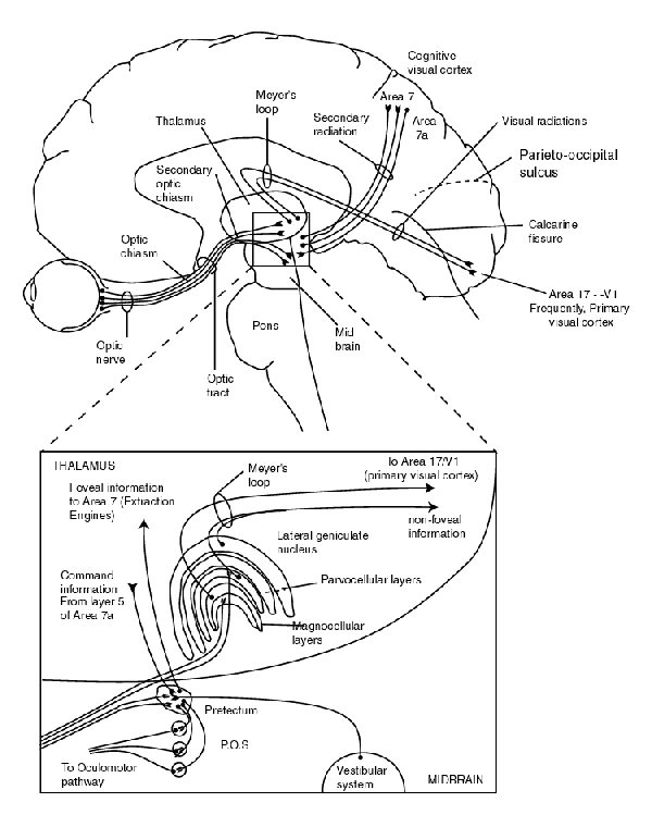

A larger scale version appears in Chapter 15 and is available for download in the Download Files area reached from the Site navigation bar.
The thalmic and mid-brain areas are shown in expanded view for clarity.
The solid signal paths represent afferent signal paths associated with the sensory neural system. Note the two distinct optic chiasms. The second chiasm separates the foveola from the non-foveola signals.
Note the two distinct signal radiations directed to the cortex. The "primary radiation" (historically named based on morphology) delivers the non-precision visual signals to area 17 of the cortex. The "secondary radiation" defined more recently through electrophysiology delivers precision visual signals to the higher order feature extraction engines of the cortex located near areas 5 and 7.
There is a counterpart to Meyer's loops consisting of the non-myelinated portions of the ganglion cells radiations within the retina.
The complete discussion of this figure is found in Section 15.2 of Chapter 15.
Return to the website home page