
The Nagel Anomaloscope was first developed in the early 1900's. It is an instrument designed for clinical evaluation of those with abnormal color vision after the individuals have been identified using flash cards. Very little guidance has been available on the meaning of the readings from the anomaloscope. The following material, from Chapter 18 of PROCESSES IN BIOLOGICAL VISION, provides a short guide in this area.
The upper frame of the figure applies to those with faulty long wavelength color vision. These people have difficulty discriminating between shades of green and shades of red. The Nagel Anomaloscope (Type I) has two controls. One control varies the amount of yellow light a subject requires to match a given mixture of red and yellow-green light. The yellow-green light is often spoken of as green for historical reasons. The second control sets the ratio of red to green light. The various lights are derived from a spectrometer and spectrally are very narrow band sources.
The subject with normal color vision always exhibits a small area of matching as indicated by the black dot in the figures. There is a small percentage of the population that have difficulty discriminating between the reds and the greens. In the worst cases, they can not discriminate between them at all except by ancillary cues (one is normally brighter than the other as in a stop signal). There is an even smaller number in the population that have difficulty discriminating between the violets and the yellows.
The horizontal dashed line is associated with subjects exhibiting poor long wavelength chrominance discrimination or rendition, but fully functional spectral detection capability. These subjects invariably match a given intensity of yellow light to a band of red green ratios. The center of this band is called the match mid point (MMP) and the range of the matches is called the match range (MR). If the MMP is not centered on the black dot, the subject exhibits an MMP error measured by the deutranomal bias. The MR can be determined using the red-green scale. Note that some deutranomalous subjects do not include the normal match point within their match range. On the other hand, a complete deutranope has an MR that includes the entire horizontal axis. His MR always includes the MMPof the color normal.
The protanope has lost all ability to sense the long wavelength or red portion of the spectrum. The locus of his vision on the anomaloscope is indicated by the complete length of the sloping line that merges with the deutranope line on the left. The matches made by the protanope never include the matches made by the color normal. For the protanomalous subject, his MR and MMP are found along intermediate lines between the deutranope and the protanope.
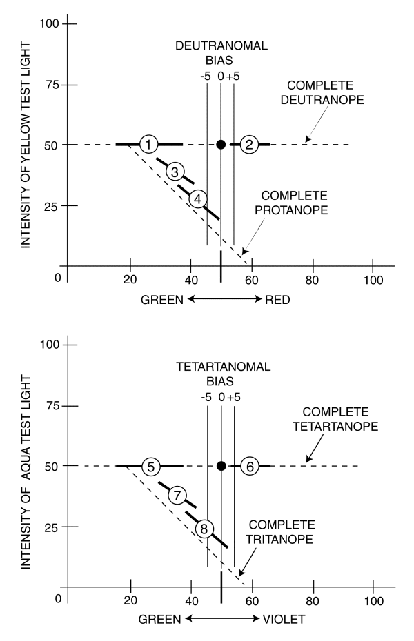
The type II Nagel Anomaloscope can be reconfigured to test those with short wavelength deficiencies. The yellow test light is replaced by an aqua test light and the red and green are replace by violet and blue-green instead. The horizontal scale is frequently marked blue-green or blue-yellow for historical reasons.
The horizontal dashed line in the lower frame is associated with subjects exhibiting poor short wavelength chrominance discrimination or rendition, but fully functional spectral detection capability. These subjects invariably match a given intensity of aqua light to a band of violet-yellow ratios. The center of this band is called the match mid point (MMP) and the range of the matches is called the match range (MR). If the MMP is not centered on the black dot, the subject exhibits an MMP error measured by the tetartanomal bias. The MR can be determined using the violet-yellow scale. Note that some tetartanomalous subjects do not include the normal match point within their match range. On the other hand, a complete tetartanope has an MR that includes the entire horizontal axis. His MR always includes the MMP of the color normal.
The tritanope has lost all ability to sense the short wavelength or violet portion of the spectrum. The locus of his vision on the anomaloscope is indicated by the complete length of the sloping line that merges with the tetartanope line on the left. The matches made by the tritanope never include the matches made by the color normal. For the tritanomalous subject, his MR and MMP are found along intermediate lines between the tetartanope and the tritanope.