
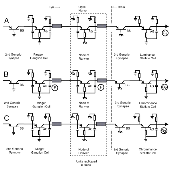
The following figure is presented in two parts. The first part shows the overall circuit diagram with the optic nerve shown in condensed form. The second part shows the optic nerve portion of the visual system with the Nodes of Ranvier replicated a number of times as required by the length of the nerve bundle.
Both of the following figures are correct for the general case of large signals. Simpler variants are available for the medium and small signal cases.
..
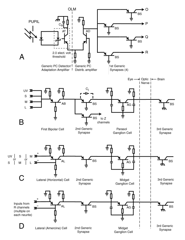
A shows the generic photoreceptor cell. The passive Outer Segment is shown on the left. It is connected acoustically to the base terminal of the adaptation amplifier, type AT, as shown by the dotted box. It is the nonlinear aspect of the impedance in the collector circuit of the adaptation amplifier and the capacitance Ca that defines the dynamic characteristics(light adaptation and color constancy) of the adaptation amplifier.
The generic output distribution amplifier, type AD,is shown in the middle with its pedicel branching to accommodate four separate generic synapses. These synapses represent symbolically the connection to the four signaling channels within the visual system, the achromatic luminance channel, R, and the three chrominance channels, O, P & Q.
Note the location of the Outer Limiting Membrane (OLM), a biological insulator electrically separating the ultimate ground connections of the two amplifiers.
B shows the generic luminance channel, R. The inputs are from each of the functional spectral channels. These signals are summed (in logarithmic space) by the bipolar cell and passed to one or more generic synapses. The most important synapse connects to a parasol ganglion cell that encodes the signal and transmits the encoded signal over the optic nerve to the 3rd genaric synapse located within the brain. A second synapse may connect to an amercine cell associated with a lateral cell (generally called an amercine cell) associated with an appearance channel
C shows the alternate case for the chromatic channels, O, P or Q. In each case, one pair of the input signals shown are subtracted (in logarithmic space) by a lateral cells (generally called a horizontal cell). The signal is then passed through a second generic synapse to a midget ganglion cell. This cell encodes the signal for transmission (projection) over the optic nerve to a 3rd synapse located within the brain.
D shows the case of the signals going to the Z channels in B (above). The signals from two different luminance channels are subtracted (in logarithmic space) and passed through a 2nd generic synapse to a midget ganglion cell for encoding and transmission to a 3rd generic synapse in the brain. The encoding is of the same type as used in C (above)
..
This figure shows three separate and distinct signal paths. The upper path is equivalent to the luminance or R channel of the above figure. The output of this channel is a monopolar electrical signal G1 representing the brightness of the scene.
The second path represents any one of the three potential chrominance channels (O, P or Q). The output of these channels are all electrically bipolar and represent the signals (in quadrature) describing the perceived color of the scene.
The lower path represents any of the appearance channels (type Z). The output of these channels are also electrically bipolar signal.
The number of Nodes of Ranvier present along any of the above signal paths is a function of the length of the path. In each of these cases, the Nodes of Ranvier operate as signal regeneration amplifiers. They do not merely amplify the received signal. They regenerate the signal to its original height and to a similar, but not necessarily identical, shape.